What your dentist does with your CBCT scan
The 3D scan on your first day is where the plan becomes real. Here is what your dentist is looking at, and what to ask.
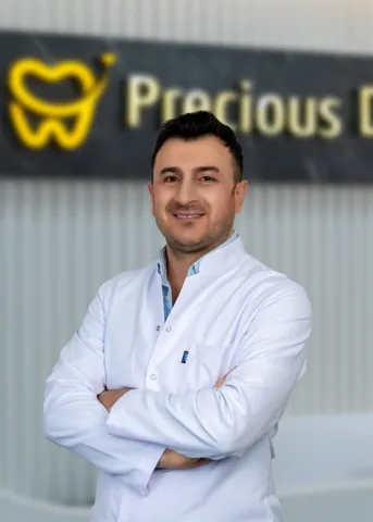 Medically reviewed by Dt. Hüseyin Yavuz Özbek, implant surgeon · Updated October 2026
A CBCT scan is a 3D X-ray of your jaws. Your dentist uses it to measure the height and width of bone where implants would go, find the nerve in the lower jaw and the sinuses in the upper jaw, check roots and infections, and plan the position, angle and size of each implant. Ask them to show you the scan and explain what changed from your first plan.
The panoramic X-ray you sent from the UK is flat. It shows your teeth and jaws from the front, but not how wide the bone is, or exactly where the nerve runs. The CBCT scan on your first day in Antalya adds the third dimension. For implants, it is the most important image your dentist will look at.
Taking the scan
You stand or sit while a machine rotates around your head for under a minute. Keep still. It does not hurt. Remove earrings, glasses and removable dentures first.
What your dentist looks at
Bone height and width
An implant needs enough bone around it. The scan lets the dentist measure, in millimetres, how tall and wide the bone is at each possible implant site.
The nerve in the lower jaw
A nerve runs through the lower jaw and gives feeling to the lip and chin. Your dentist plans implants to stay a safe distance from it.
The sinuses
Above the upper back teeth are the air spaces of the sinuses. If the bone below them is thin, the dentist may plan a sinus lift or angle implants forwards.
Roots, infections and cysts
The scan can show infection at the tips of roots, cracks and cysts that a flat X-ray misses. This can change whether a tooth is saved or removed.
From scan to plan
Many dentists plan implants on the scan with software: they place virtual implants, choose the length and width, and check the angle against the teeth that will go on top. For full-arch cases, this is how the number and position of implants is decided.
At Precious Dental, full-arch cases are planned from a 3D scan, and you are told in writing how many implants you need, which brand and why.
What to ask when they show you
- How much bone do I have where the implants will go?
- Is there anything here that changes the plan I was sent?
- Will I need a graft or sinus lift? Why?
- Where is the nerve, and how close will the implants be?
- Are there any teeth you could save that you planned to remove, or the other way round?
When the plan changes
It is common for the scan to change something: one more implant, one fewer, a graft, a tooth that can be saved. A good dentist will show you why on the screen. A big change with no clear explanation is a reason to pause. Read warning signs in the chair.
Keep your scan
Ask for a copy of the scan files. They are part of your records and a UK dentist may need them years from now. Read guarantees and records.
Reading the screen together
When your dentist shows you the scan, it can look like grey clouds. Ask them to point out a few landmarks: your teeth or gums, the outline of the jawbone, the dark channel of the nerve in the lower jaw, and the dark air space of the sinus above the upper back teeth. Once you can see those, the plan makes sense. You can see where the implants will sit and why the dentist chose that position.
Slices and 3D views
The scan can be viewed as a 3D model or as thin slices through the jaw. Dentists often use cross-section slices to measure bone width at each implant site, because the width is what a flat X-ray cannot show. If your dentist says “the bone is narrow here”, ask to see the slice. It is the clearest way to understand why a graft or a different implant position is suggested.
Guided surgery
Some dentists use the scan to make a surgical guide: a plastic template that fits over your teeth or gums and guides the drill to the planned position and angle. Others plan on the scan and place implants freehand using the plan as a map. Both approaches are used by experienced implant dentists. Ask which your dentist uses and why.
Scans for veneers and crowns
For cosmetic work, the important scan is often a digital scan of the teeth, taken with a small camera in the mouth, rather than a CBCT. It gives the lab an exact model to design on. A CBCT may still be useful if a tooth has an old root canal or a suspected infection.
A scan as a baseline
Your first scan becomes a baseline. If you have a problem years later, comparing a new X-ray with the original helps any dentist see what has changed around the implants. That is one more reason to keep the files safe.
What the scan can show that changes plans
| Finding | What it may mean for your plan |
|---|---|
| Narrow bone | Narrower implant, graft, or a different position |
| Low bone under the sinus | Sinus lift, shorter implant, or angled implants |
| Nerve close to the planned site | Shorter implant or a different site |
| Infection at a root tip | Treat the infection, or extract and wait before an implant |
| Healthy root you were told to lose | A tooth may be saved with root canal treatment |
| Thickened sinus lining | Possibly a sinus problem to check before surgery |
The sinus, in plain words
The maxillary sinuses are air spaces in the cheekbones, above the upper back teeth. The NHS explains that sinusitis is swelling of the sinuses, usually after a cold, and usually gets better on its own. If you have frequent sinus problems, tell your dentist. A sinus lift lifts the floor of the sinus to make room for bone, and a dentist will want the sinus to be healthy before doing it. Read health conditions to tell your dentist.
Radiation and scans
A CBCT uses X-rays. Dentists use it when the 3D information will change the plan, as it usually does for implants. If you have had a recent scan elsewhere, bring the files: a dentist may be able to use them. If you are or might be pregnant, tell the clinic before any X-ray.
Second opinions from the scan
Because the scan is a file, it can be shared. If the plan changes a lot after your scan and you are unsure, you can ask for a copy and show it to another dentist, in Antalya or at home. A careful dentist will not mind. Read warning signs in the chair.
Scan questions checklist
- Show me the bone at each implant site.
- Where is the nerve and the sinus?
- Is any graft needed, and how sure are you?
- Does anything here change the plan or price?
- Can I have the files before I leave?
Our implant surgeon, Dt. Hüseyin Yavuz Özbek, plans full-arch cases from a 3D scan. See the other implant dentists in Antalya.
If you have had implants before
If you already have implants elsewhere in your mouth, the scan also shows them. Bring any records you have of their brand and size. Your dentist can check how they look and plan new implants around them. If an old implant has bone loss around it, they may suggest dealing with that first. Read guarantees and records.
Seeing the result after surgery
Many dentists take an X-ray after implant surgery to check the final positions. Ask to see it next to the plan on your scan. It is reassuring to see the implants where they were planned, and the image becomes part of your records.
Common questions
What is a CBCT scan?
Cone beam computed tomography: a 3D X-ray of the jaws and teeth. It takes less than a minute while you stand or sit still.
Is a CBCT scan safe?
It uses X-rays, at a higher dose than a normal dental X-ray but much lower than a medical CT. Dentists use it when the 3D information is needed, as it is for implants.
Can I get a copy of my scan?
Yes. Ask for the files before you leave. A UK dentist may need them later.
Why did my plan change after the scan?
Because the scan shows bone, nerves and sinuses that a flat X-ray cannot. Ask your dentist to show you exactly what changed.
Sources
- NHS: treatment abroad checklist
- Oral Health Foundation: going abroad for dental treatment
- NHS: sinusitis
Checked October 2026. Where we give our own view as a clinic, we say so.
Send your question to a Precious Dental dentist
Send an X-ray or a few photos. A Precious Dental dentist will reply with a written plan and price. Ask us who would treat you at each stage. No obligation.